Diagram Of The Muscles In The Forearm / Tendons In Right Hand 7. Muscles Of The Forearm And Hand ... : 2, ulna, 3, biceps muscle;. Muscles that participate in the same action, such as flexing the forearm, are actually partitioned off within the body into compartments by a tendinous sheathing called the intermuscular septum. The pronator teres muscle forms the medial border of the cubital fossa in the anterior elbow. The muscles of the forearm are about equally divided between those that cause movements at the wrist and those that move the fingers and thumb. Start studying muscles of the forearm. The muscles in the posterior compartment of the forearm are commonly known as the extensor muscles.
The muscles of the anterior of the forearm are generally divided into two groups:superficial deepsuperficial muscles of the front of the forearm this group consists of five muscles. Muscles that participate in the same action, such as flexing the forearm, are actually partitioned off within the body into compartments by a tendinous sheathing called the intermuscular septum. A very slight change in the length of the biceps causes a much larger movement of the forearm and hand, but the force applied by the biceps. In these diagrams, the brachioradialis muscle is indicated. 2, ulna, 3, biceps muscle;
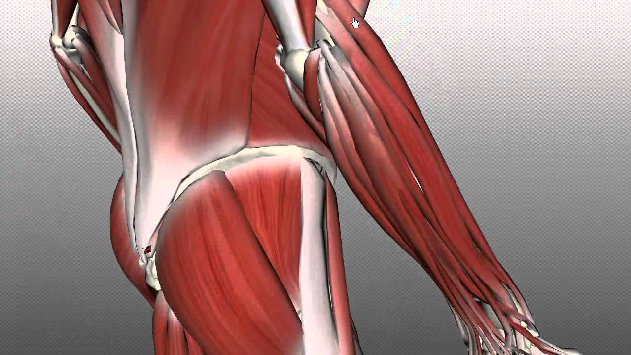
Posterior View of the Superficial Muscles of the Arm ... from etc.usf.edu In the posterior compartment, you can separate the muscles into a superficial layer and a deep layer. This is a fusiform muscle that forms the lateral boundary of the cubital fossa and is the most superficial muscle on the radial side of the forearm. The flexor pollicis longus is situated on the radial side of the forearm, lying in the same plane as the preceding. This muscle is part of muscle anatomy master class. The forearm is the region of the upper limb between the elbow and the wrist. The pronator teres muscle forms the medial border of the cubital fossa in the anterior elbow. Diagram of the muscles of the arm in action. The muscles in the posterior compartment of the forearm are commonly known as the extensor muscles.
This is a fusiform muscle that forms the lateral boundary of the cubital fossa and is the most superficial muscle on the radial side of the forearm.
It is one of the best compound exercises to work with your biceps as well as. Superficial muscles of the posterior forearm: The superficial layer contains four of these on the next diagram we will indicate the intermediate layer of anterior compartment of forearm. Inflammation of this region caused by repetitive. In these diagrams, the brachioradialis muscle is indicated. Remembering the action of each one can be quite difficult. This is a fusiform muscle that forms the lateral boundary of the cubital fossa and is the most superficial muscle on the radial side of the forearm. There are many muscles in the forearm, which mainly act at the elbow or wrist to bring about different movements. The muscles in the posterior compartment of the forearm are commonly known as the extensor muscles. 4, attachment… the muscles of the back forearm. The forearm is the region of the upper limb between the elbow and the wrist. Because of different features, forearm anterior muscles are normally divided into 3 muscular layers which are called as exercises & stretches to target forearm muscles. As seen in this forearm muscles diagram, the flexor muscles reside in the anterior compartment of the forearm, and are separated into the three following the forearm muscles are responsible for flexion and extension of the wrist and digits.
2, ulna, 3, biceps muscle; The accompanying muscle diagram reveals the muscles' positions beneath the surface. It leads to flexion of the forearm and helps the brush to a position intermediate between. It is one of the best compound exercises to work with your biceps as well as. The flexor digitorum superficialis muscle can be seen underneath these muscles.
Forearm Muscles Part 2 - Posterior (Extensor) Compartment ... from i.ytimg.com The anterior forearm muscles are divided into 3 muscular layers; A deep layer, intermediate layer and superficial layer. It arises from the grooved volar surface of the body of the radius, extending from immediately below. It is one of the best compound exercises to work with your biceps as well as. Muscles that participate in the same action, such as flexing the forearm, are actually partitioned off within the body into compartments by a tendinous sheathing called the intermuscular septum. The muscles of the upper arm are responsible for the flexion and extension of the forearm at the elbow joint. Start studying muscles of the forearm. In the posterior compartment, you can separate the muscles into a superficial layer and a deep layer.
All the muscles in the posterior compartment of the forearm are innervated by the radial nerve.
The muscles of the upper arm are responsible for the flexion and extension of the forearm at the elbow joint. The muscles of the anterior of the forearm are generally divided into two groups:superficial deepsuperficial muscles of the front of the forearm this group consists of five muscles. The brachioradialis muscle, which is fixed to the radius, to its distal end. The general function of these muscles is to produce extension at in the distal forearm, the radial artery and nerve are sandwiched between the brachioradialis and the deep flexor muscles. Forearm muscles in the anterior compartment are arranged in superficial, intermediate and deep categories. Learn and reinforce your understanding of muscles of the okay, before we start, it is important to know that, even though some of the muscles of the forearm attach proximally to the humerus, they still belong. Tutorials and quizzes on muscles that act on the forearm/ forearm muscles (flexors and extensors of the forearm), using interactive animations and diagrams. Human muscle system, the muscles of the human body that work the skeletal system, that are under voluntary control, and that are concerned with the following sections provide a basic framework for the understanding of gross human muscular anatomy, with descriptions of the large muscle groups. Muscles of the forearm videos, flashcards, high yield notes, & practice questions. Superficial muscles of the posterior forearm: In fact, there is another muscle grouped underneath it named extensor carpi radialis longus. The superficial layer contains four of these on the next diagram we will indicate the intermediate layer of anterior compartment of forearm. Try labeling diagrams and worksheets as additional learning aids.
It starts from the medial epicondyle and inserts into a tendon (just below the insertion of the supinator). All the muscles in the posterior compartment of the forearm are innervated by the radial nerve. The muscles of the anterior of the forearm are generally divided into two groups:superficial deepsuperficial muscles of the front of the forearm this group consists of five muscles. The flexor pollicis longus is situated on the radial side of the forearm, lying in the same plane as the preceding. Click here for access to the full anatomy glossary.
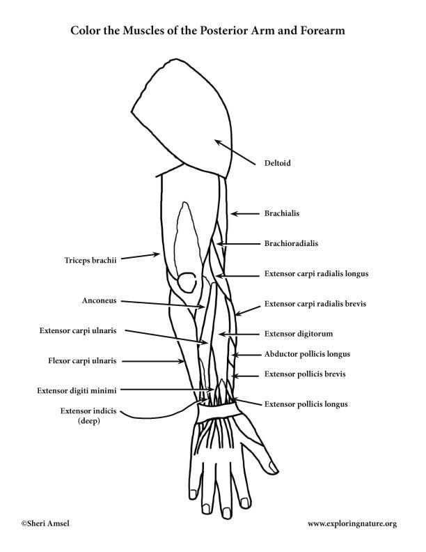
Muscles of the Arm and Forearm (Posterior) - Coloring Page from www.exploringnature.org The forearm is the region of the upper limb between the elbow and the wrist. Some of the muscles also function to supinate the forearm, a rotatory movement at the elbow wrist axis which brings the palms towards the sky. As a result musculoskeletal disorders appear 12. Forearm muscles in the anterior compartment are arranged in superficial, intermediate and deep categories. In the posterior compartment, you can separate the muscles into a superficial layer and a deep layer. Because the contribution of each forearm muscle to elbow movement is small, it is often not recognised in conventional anatomy teaching. This human anatomy diagram with labels depicts and explains the details and or parts of the muscles in the forearm. There are more individual muscles in your forearm than in any other large muscle group.
4, attachment… the muscles of the back forearm.
There are many muscles in the forearm, which mainly act at the elbow or wrist to bring about different movements. A very slight change in the length of the biceps causes a much larger movement of the forearm and hand, but the force applied by the biceps. Superficial muscles of the posterior forearm: I made an entire tutorial dedicated to drawing the forearms with anatomical detail, it can be fond here. This layer contains only one muscle, the flexor digitorum. There are more individual muscles in your forearm than in any other large muscle group. Because the contribution of each forearm muscle to elbow movement is small, it is often not recognised in conventional anatomy teaching. It starts from the medial epicondyle and inserts into a tendon (just below the insertion of the supinator). Human anatomy diagrams and charts show internal organs, body systems, cells, conditions, sickness and symptoms information and/or tips to ensure one lives in good health. In these diagrams, the brachioradialis muscle is indicated. Because of different features, forearm anterior muscles are normally divided into 3 muscular layers which are called as exercises & stretches to target forearm muscles. A deep layer, intermediate layer and superficial layer. The flexor pollicis longus is situated on the radial side of the forearm, lying in the same plane as the preceding.


0 Komentar